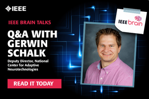
Gerwin Schalk, Ph.D., Deputy Director of the National Center of Adaptive Neurotechnologies, was an early adopter of electrocorticography (ECoG) for the study of brain function and neurological disorder therapies. He will be a keynote speaker at NeuroCAS, a collaborative workshop to explore the challenges of neurotechnology held 21-22 October in Cleveland, Ohio, directly following BioCAS 2018. IEEE Brain recently spoke with him about the opportunities ECoG presents for both research and clinical applications.
Why has interest in using ECoG for research gained popularity recently?
Gerwin Schalk: Interest has grown because of the tremendous opportunities ECoG provides both for basic research and for clinical applications. For example, we know that even simple functions require interactions of many different areas of the brain and if you record only from a few neurons in one small part of the brain you may be getting fairly good information about a specific action such as moving your finger, but you will not know anything about the process leading up to that decision. If you want to understand how information is transferred from one area of the brain to another, you need to gather signals from multiple locations in order to understand that process as a whole. ECoG is really the only method that offers high spatial resolution, high resolution in time, and provides measurements across large areas of the brain. There’s simply no other imaging technique that can do this at this time.
Where do you see opportunities for ECoG in both basic research and clinical applications?
Gerwin Schalk: The basic research opportunity is accelerated by techniques specific to ECoG that essentially take better advantage of the information these high-resolution images provide. If you have high spatial resolution and if you study each activity at 100 locations independently resulting in 1000 measurements per second—that’s 100,000 numbers a second. If you want to study the interactions between those, that’s 100,0002. Therefore, you need to define a way to extract from those many numbers the activity of interest, preferably something that teaches you about how the brain actually does something. There are a lot of computational challenges and those challenges are now starting to be addressed by researchers; that’s really where the opportunities lie in the basic research domain today.
The clinical opportunities center on interfacing with the brain in one way or another for a beneficial medical purpose. In these use cases, what you really need are stable measurements and a stable interface with the brain because it needs to do something in real time and, in the case of communication, on pretty rapid time scales. Ideally, you would need an electrical method that at least in principle can support rapid interactions, and for this reason both the EEG and the single neuron recording techniques have problems. The EEG technique is simple and safe and non-invasive but the signals that you get are not very good—the noise can often be so large that for some time all you may see is noise and you aren’t able to measure anything useful. In contrast, the single neuron recording technique can effectively measure activity from individual neurons, but the brain doesn’t really like those tiny implants and encapsulates them, and after some time you stop being able to hear activity from these neurons. These approaches may be fine for basic research if it’s limited in time and scope but if you really want to help someone who is disabled, it’s not viable if your device stops working due to noise levels, or if it degrades after six months.
ECoG is the only method that when you implant it, it’s going to give you very robust signals for years. There’s strong scientific evidence now where people have implanted in monkeys and the ECoG devices have provided ongoing quality signals. That’s something of major significance when you consider taking this out of the laboratory and helping people that have neurological disorders. The fact that it provides a stable interface–that’s the tremendous opportunity on the clinical side.
What are the challenges in employing effective ECoG devices?
Gerwin Schalk: The challenge for basic research is that we typically only get recordings from the cortical surface. Because we are recording only on the lateral surface and not inside the folds and even less so inside the brain then we still have a very sparse and incomplete image. What we would really want is to have the same kind of capacity, resolution, and signal quality as these ECoG electrodes but somehow magically have that information from every place in the brain, which essentially requires a completely new technology that’s not yet conceived.
Another challenge is that interpreting the signals is more difficult the better the signal quality, and that actually goes into a really deep sense of that meaning. It’s become obvious that the procedures we use currently to get information are fundamentally sub-optimal and basically flawed. To be efficient and effective, you need to have some idea what you are trying to extract so that you can write the signal processing techniques to do this extraction. The problem is that we don’t know what the brain does and what kind of brain signatures we should be looking for and ultimately, the ways in which we extract them are essentially violating the actual nature of these signatures. We see things we think are there but they really aren’t there—they’re just an artifact of the signal extraction, for example. What we really need is a better understanding of how neurons create electrical activity, and how that electrical activity somehow gives rise to the signal signatures that we actually measure with these electrodes. In theory it’s simple biophysics, but what we still don’t know is how all these neurons are connected and how they are geometrically associated with each other so we really can’t use physics to simulate and to understand the signals that we are actually measuring. Ultimately, there are fundamental unsolvable challenges as well as some solvable challenges here because we now know a little more than nothing and that information can be used to provide the basis for new methods for signal extraction in the future.
On the clinical side, there’s a formidable challenge, which is essentially a financial problem. If you are hoping to develop a new invasive device to use for people who have a certain disorder there is going to be trial and error, and every “try” costs about US$100 million. Yet, the reason we still need to try different devices regardless of the fact that our approach may be radically biased is that we are limited by the lack of a comprehensive model of the brain. If we understood the brain then we could pinpoint what’s wrong and we could design a device in a particular way to accomplish a particular thing. But because we are limited in our understanding, we may be guessing for many years and likely there won’t be enough funding available. However, I think the best we can do is continue to increase our understanding of the brain and then take that knowledge and use it to inform the design of new devices and protocols, ultimately creating more efficient and effective therapies.


